Total Knee Arthroplasty:
Total Knee Replacement
Anatomy
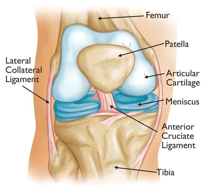
The knee is the largest joint in the body and having healthy knees is required to perform most everyday activities.
The knee is made up of:
- The lower end of the thighbone (femur)
- The upper end of the shinbone (tibia)
- The kneecap (patella)
The ends of these three bones are covered with articular cartilage, a smooth substance that protects the bones and enables them to move easily within the joint.
The menisci are located between the femur and tibia. These C-shaped wedges act as shock absorbers that cushion the joint.
Large ligaments hold the femur and tibia together and provide stability. The long thigh muscles give the knee strength.
All remaining surfaces of the knee are covered by a thin lining called the synovial membrane. This membrane releases a fluid that lubricates the cartilage, reducing friction to nearly zero in a healthy knee.
Normally, all of these components work in harmony. But disease or injury can disrupt this harmony, resulting in pain, muscle weakness, and reduced function.

Cause

The most common cause of chronic knee pain and disability is arthritis. Although there are many types of arthritis, most knee pain is caused by just three types: osteoarthritis, rheumatoid arthritis, and posttraumatic arthritis.
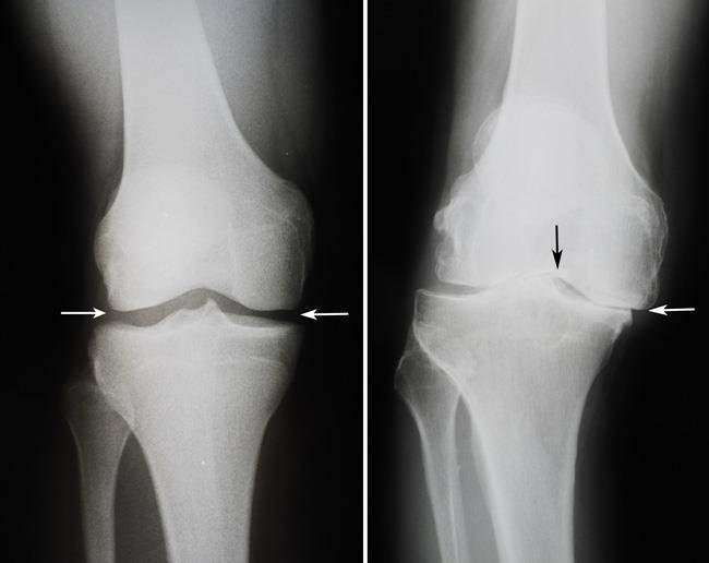
- Osteoarthritis. This is an age-related wear-and-tear type of arthritis. It usually occurs in people 50 years of age and older, but may occur in younger people, too. The cartilage that cushions the bones of the knee softens and wears away. The bones then rub against one another, causing knee pain and stiffness.
- Rheumatoid arthritis. This is a disease in which the synovial membrane that surrounds the joint becomes inflamed and thickened. This chronic inflammation can damage the cartilage and eventually cause cartilage loss, pain, and stiffness. Rheumatoid arthritis is the most common form of a group of disorders termed “inflammatory arthritis.”
- Posttraumatic arthritis. This can follow a serious knee injury. Fractures of the bones surrounding the knee or tears of the knee ligaments may damage the articular cartilage over time, causing knee pain and limiting knee function.
The Orthopaedic Evaluation
An evaluation with an orthopaedic surgeon consists of several components:
- Medical history. Your orthopaedic surgeon will gather information about your general health and ask you about the extent of your knee pain and your ability to function.
- Physical examination. This will assess knee motion, stability, strength, and overall leg alignment.
- X-rays. These images help to determine the extent of damage and deformity in your knee.
- Other tests. Occasionally, blood tests or advanced imaging, such as a magnetic resonance imaging (MRI) scan, may be needed to determine the condition of the bone and soft tissues of your knee.
Your orthopaedic surgeon will review the results of your evaluation with you and discuss whether total knee replacement is the best method to relieve your pain and improve your function. Other treatment options — including medications, injections, physical therapy, or other types of surgery — will also be considered and discussed.
In addition, your orthopaedic surgeon will explain the potential risks and complications of total knee replacement, including those related to the surgery itself and those that can occur over time after your surgery.


